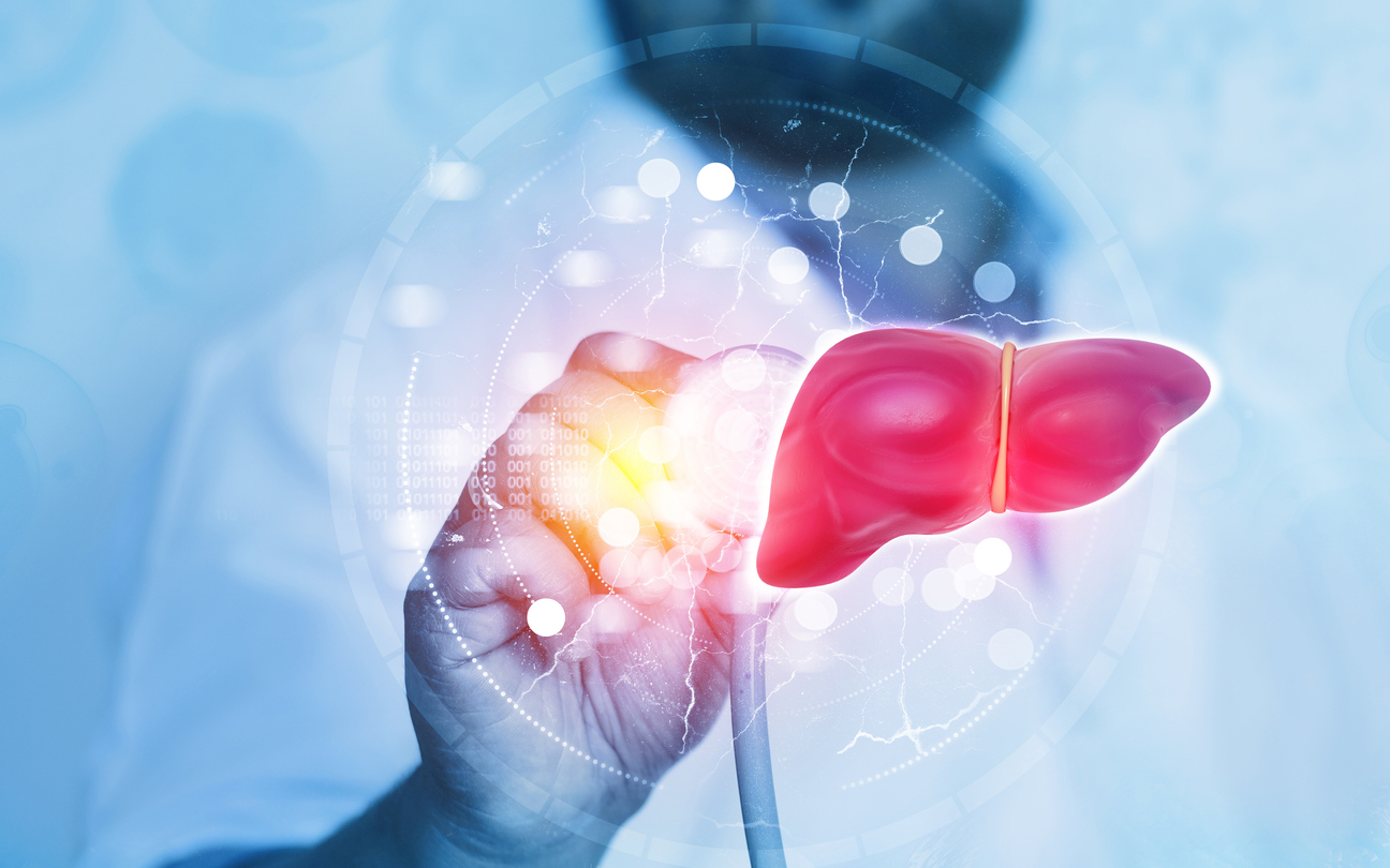
The liver is a reddish-brown colored organ and gland in the human body located on the right side, under your right ribs and beneath your right lung. The largest organ in the body, some of its primary functions are filtering blood and toxins, producing bile, helping blood clot, and aiding the metabolism of proteins, carbohydrates, and fats. Essentially, everything you eat is broken down by the liver. The liver also stores glycogen, makes chemicals and proteins and rids the body of old blood cells.
The liver has a larger right lobe and a smaller left lobe.
The cells in the liver are primarily of a type called hepatocytes. It has other cell types that line its blood vessels and the bile ducts (which carry bile to the gallbladder or intestines).
Depending on which type of cells develop into tumors, they are classified as benign or malignant. Each liver tumor requires its diagnosis.
Primary liver cancer: The origin of cancer in the liver is called primary liver cancer. The main types of primary liver cancers are:
Hepatocellular carcinoma (HCC) is the most common form of liver cancer. HCC typically begins as a single tumor and develops in the hepatocytes, and cancer grows and spreads to other areas in the liver in its later stages. HCC is usually a consequence of liver cirrhosis, a condition caused by excessive alcohol consumption that scars liver tissue and hardens it, severely hampering its functions. Cirrhosis can also be caused by Hepatitis B or C virus, which attacks the liver.
Another type of HCC has several small cancer growths in the liver, also triggered by cirrhosis.
Fibrolamellar carcinoma: A relatively uncommon sub-type of cancer, it affects people in their younger years of the 20s and 30s and is not connected to liver damage.
Intrahepatic cholangiocarcinoma (Bile Duct Cancer): Cholangio is a term for bile ducts, the thin channels that carry bile to the stomach or intestines. This type of cancer can begin in bile ducts inside or outside the liver.
Angiosarcoma is a rare and rapidly spreading liver cancer in the cells lining the liver's blood vessels.
Hemangiosarcoma: Another rare form of liver cancer, it begins in the cells lining the liver's blood vessels and spreads rapidly, making it challenging to treat.
Secondary liver cancer (Metastatic Liver Cancer): This category of cancer does not originate in the liver but spreads to it from another area in the body; hence, it is secondary cancer.
When the cells in the liver change their DNA, it triggers unpredictable cell behavior that may result in uncontrolled cell division, leading to the formation of tumors. Several factors increase the risk of this cell response:
Chronic Hepatitis B or C Virus infection: People infected with HBV or HCV are at high risk of developing liver cancer. The HBV virus causes severe liver inflammation, while HCV can trigger cirrhosis, and both can lead to liver cancer.
Cirrhosis: This condition leads to scarring of the liver tissue, which prevents the liver cells from functioning normally. Excessive alcohol consumption and hepatitis infections can be critical triggers of cirrhosis.
Diabetes. Uncontrolled diabetes increases sugar levels in the body, putting extreme pressure on the liver to metabolize it. If this goes unchecked, it can trigger cancer as the liver cells get damaged.
Nonalcoholic fatty liver disease: Obesity causes the liver to begin storing extra fat, and if it is not treated, it can lead to cancer due to cirrhosis. The presence of high levels of triglycerides or LDL ("bad") cholesterol is also a contributing factor. Unwanted elements in the liver, such as fat, sugar, and non-metabolized substances such as excessive alcohol, etc., can severely strain the liver cells causing them to break down. It can trigger malignancy as cell behavior is altered due to severe performance stress.
A subtype of nonalcoholic fatty liver disease is NASH or nonalcoholic steatohepatitis, indicating inflammation and liver cell damage.
Family history: If an immediate family member, such as a sibling or a parent, has primary liver cancer, it increases the chances of getting liver cancer.
Proximity to aflatoxins. A carcinogenic fungus, aflatoxins are found growing on foods such as wheat, soybeans, nuts, etc., and prolonged exposure can cause liver cancer.
Smoking: It can increase the risk of liver cancer.
Hereditary hemochromatosis: This condition makes the person absorb excessive iron from foods which, when stored in the liver, can damage the cells and cause cirrhosis, potentially triggering cancer.
Excessive alcohol intake: Prolonged alcohol consumption damages the liver and can lead to cancer.
At the onset of primary liver cancer, few symptoms alert you to its presence. But as the disease progresses, you may notice some or several of the following signs:
Unintentional weight loss and loss of appetite: The liver is critical to digestion and appetite. When cancer sets in, it cannot carry out its normal functions, leading to poor appetite and unwanted weight loss.
Nausea and vomiting: Due to a malfunctioning liver, the body ends up with high levels of calcium, which leads to nausea, and muscle problems.
Weakness/fatigue: The body is not getting its required nutrients due to loss of appetite, and the liver's many functions to keep it healthy and toxins-free are impaired, leading to weakness.
Jaundiced look: When the liver cells get cancerous, their reduced function increases the presence of bilirubin, leading to a condition called Hyperbilirubinemia. Yellow color in the eye whites, a yellow tint on the skin, pale stools, and dark yellow urine characterize it. Bile duct cancers can bring on early symptoms of jaundice.
Fever, engorged veins on the belly, and unexplained bruising or bleeding.
Hepatic encephalopathy: The liver is a clearinghouse for toxins, and when it gets damaged, it can no longer do the job effectively. So, toxins build up, and when they reach the brain, they can mess with its functions leading to fatigue, poor focus, sleepiness, and disorientation.
Hard lump under rib cage: It is caused due to the enlarged liver.
Pain in the upper abdomen: The growing tumorous mass in the liver expands in size, pressing on the abdomen and causing pain. The pain may also be due to the blood backing up in the vessels and seeping into the stomach, leading to swelling.
Tumor-produced hormones: Cancerous tumors produce hormones that can trigger diseases in other body parts. Remember, a healthy liver and a healthy body control the role of hormones, but a diseased liver cannot. Some conditions due to malignant tumor-produced hormones are Hypoglycemia or low blood sugar, gynecomastia or breast enlargement, testicular shrinkage, increased red blood cells (Erythrocytosis), and high cholesterol.
Liver cancer staging has different systems.
The United States uses AJCC (American Joint Committee on Cancer) TNM system, which
considers the following factors:
o How large is the tumor (T)?
o Has it spread outside the pancreas affecting the nearby lymph nodes (N)?
o Has it metastasized (spread to other body parts) (M)?
The staging is based on various tests, examinations, biopsies, and imaging tests (ultrasound, CT, or MRI scan, for example). TNM has four stages: I to IV.
Stages IA and B: These stages reveal the presence of a single small-sized localized tumor or a
tumor larger than 2cms.
Stage II: One tumor larger than 2cm reaching the blood vessels or more tumors about 5 cm but not spread to lymph nodes or beyond.
Stages III A and B: There are more tumors; one is larger and localized, and in Stage B, one tumor has invaded the liver's major vein. The malignancy has not reached the lymph nodes or beyond.
Stages IV A and B: One or several tumors reach nearby lymph nodes and, in Stage B, spread to organs beyond though the lymph nodes are safe.
American Hospital Dubai follows the AJCC TNM staging system.
The Barcelona Clinic Liver Cancer (BCLC) Staging System
It has five stages:
0: Initial stage: The individual is active, and the liver functions well.
A: Early stage: Active and liver not under significant stress. A tumor is asymptomatic.
B: Intermediate: Multiple tumors exist, but the individual is functioning well.
C: Advanced: Cancer is spreading, the individual is not well, but the liver is holding up.
D: Terminal: Cancer has spread, and the patient is debilitated.
Several methods are used to diagnose liver cancer, depending on the stage and spread of the disease.
Physical exam and medical history: The doctor will conduct a thorough evaluation, ask about their health and family history and perform a physical examination to check for signs and symptoms of liver cancer.
Blood tests: Doctors conduct several types of blood tests depending on what they are looking for as cancer markers:
Liver Function Tests (LFTs): These are done to see the degree of liver damage and impaired functions.
Tests to check blood clotting: One of the main functions of a healthy liver is to help the blood clot to prevent excessive bleeding. A blood clotting test assesses the degree of liver damage.
Blood chemistry tests: These tests check for volumes of certain substances, like calcium, sugar, cholesterol, etc., in the blood that can be impacted by liver cancer. A malfunctioning liver can interfere with their normal levels.
Hepatitis B and C tests: Hepatitis B virus and Hepatitis C virus can cause significant damage to the liver and cause cancer. These tests are done to assess the degree of damage.
Kidney function tests: Tests of blood urea nitrogen (BUN) and creatinine levels are often done to assess how well your kidneys are working.
Imaging tests (Ultrasound, CT, MRI, X-rays, etc.): These tests produce a picture of the liver and other internal organs that may be affected by cancer by revealing abnormalities of growth (tumors) and its spread. These tests also differentiate between benign and malignant tumors and reveal blockages in blood vessels in and out of the liver.
Liver biopsy: Liver tissue sample is extracted to be checked for malignancy using a thin needle inserted into the liver through the skin to get the tissue sample, and several methods are employed, including needle biopsy, laparoscopy, or surgically removing a sample. A cautionary note for doctors is the possibility of agitating the cancer cells with a needle, so needle biopsy is primarily done only when the patient is primed for a liver transplant.
Alpha-fetoprotein blood (AFP) test: Liver damage leads to high levels of a protein called Alpha-fetoprotein, though AFP levels can be increased due to other factors. But AFP tests during liver cancer treatment can reveal its efficacy. Typically, AFP levels decrease if the treatment works, and AFP is also used to determine if the cancer is recurring.
Angiography is an X-ray test that reveals the state of the arteries carrying blood to the liver.
The stage, grade and spread of cancer decide the treatment path. Several approaches are used to combat liver cancer:
Surgery: Early-stage liver cancer responds well to surgery, provided the liver is mainly healthy, with cancer contained in a small area. The cancerous portion and a small piece of a healthy liver are removed.
Liver transplant surgery: A donor's healthy liver replaces the damaged liver. There are regulations for a liver transplant, and the Milan criteria in transplantation medicine are widely followed for patients with cirrhosis and Hepatocellular Carcinoma (HCC). The requirements stipulate that transplant recipients must have a single HCC tumor no bigger than 5cm or two or three tumors of 3cm or less at the time of diagnosis.
There are ongoing discussions about the criteria.
Chemotherapy: This treatment uses drugs to kill or disable cancerous cells. The drugs are either orally administered or injected into the body.
Chemoembolization: Drugs are injected into blood vessels near the tumor in the liver, or the drugs are attached to small beads and injected into an artery flowing to the tumor. The beads block the blood flow to the tumor, and the drugs kill the malignant cells.
Radiation beads: Tiny beads with radiation are inserted into the liver to attack the tumor and kill it.
Tumor ablation: The word ablation means removal. The high heat generated from radio waves or laser (radiofrequency ablation, RFA) or alcohol injections (Percutaneous Ethanol Injection) is administered with an ultrasound scan to target the cancer cells precisely.
Targeted Cancer Therapy: This treatment uses drugs that hamper the molecular behavior of cancer cells, stopping cancer from growing.
Radiotherapy: High-energy rays are directed at cancer cells to thwart their growth or kill them. This treatment can be delivered from outside the body or from within, though external radiation is not usually given if the liver is highly damaged as it cannot tolerate it. Internal radiation sends a radioactive substance to the cancer cells through the hepatic artery.
Cryoablation: Extreme cold temperatures are administered to the cancer cells, freezing and killing them, using ultrasound to reach the malignant site.
Immunotherapy: The cancer patient's immune system fights the disease through advanced methods to attack the cancer cells that trick the immune system and evade detection.
Palliative care is a form of therapy that alleviates cancer pain and associated symptoms and seeks to comfort patients while they undergo treatments.

English, Urdu

Arabic, English

Arabic, English, French